
Researchers at NeuroElectronics Flanders (NERF) have published two new studies advancing our understanding of how visual information is processed in the brain. These studies reveal the complexity of the brain’s visual processing network and how it adapts to different states of arousal and behavior.
The brain is constantly processing streams of sensory input, transforming them into representations that guide perception and behavior. In the visual system, this begins with the retina, which captures the world around us and converts it into electrical signals that are sent to the brain.
“We know that different neurons in the visual system are specialized for distinct functions,” explains Dr. Vincent Bonin, professor at KU Leuven and group leader at NERF. “Some help us perceive fine details, while others process motion. What we still don’t fully understand is how these specialized functions emerge from the brain’s circuitry and how behavior modulates them. Understanding this is key to uncovering how the brain reconstructs the visual world.”
The first study, led by NERF postdoctoral researcher Xu Han and published in Current Biology, challenges the traditional “linear flow” model of visual processing. Instead of information moving step by step—from the retina to the thalamus and then to the cortex—the team found a more intricate and dynamic network.
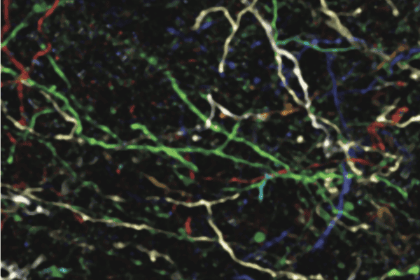
Using advanced circuit-tracing techniques and neural imaging in mice, the researchers identified specialized connectivity patterns in the brain. Some pathways selectively carry distinct types of information, while others broadcast signals more broadly.
“For instance, neurons in the pulvinar and certain layers of the cortex are finely tuned to their targets, suggesting a role in constructing detailed visual representations,” says Han. “In contrast, deeper neurons seem to ignore target specificity, broadcasting similar visual information across areas—possibly for coordinating broader brain activity.”
These findings reveal distinct roles for higher-order visual pathways and show the brain’s visual system as a highly adaptable network, capable of integrating and transmitting information in complex ways.
In the second study, published in Cell Reports, Dr. Bonin and Dr. Karolina Socha (now at UCLA) explored how the brain’s thalamus—a key relay station for visual signals—adjusts its processing depending on behavioral states. The researchers found that during quiet wakefulness, neurons in the thalamus amplify signals for back-to-front motion, a transformation that is absent under anesthesia or heightened arousal.
By imaging the activity of neurons in awake mice, the team discovered that this behavioral modulation is linked to changes in pupil size, a marker of arousal. Larger pupils coincided with stronger responses to back-to-front motion, suggesting that the thalamus integrates sensory inputs with behavioral context to prioritize certain visual stimuli.
Our findings demonstrate how the thalamus integrates behavioral context to dynamically shape visual representations, altering how motion is processed and prioritized. The results highlight an intricate interplay between sensory input and behavior, with potential implications for how the brain prioritizes visual stimuli in real-time.
Together, these studies take us closer to creating a detailed functional map of the visual system. Bonin emphasizes the importance of this work: “By understanding how different areas and cells contribute to visual processing and behavior, we can uncover the fundamental principles of brain function.”
Looking ahead, the team plans to manipulate specific pathways systematically to observe how they influence perception and behavior. This approach could help identify critical circuits for specific visual tasks, providing a roadmap for future research and potential clinical applications.
By moving beyond static brain maps, these studies pave the way for interventions to address visual and neural dysfunctions, opening new avenues for both neuroscience and medicine.
_
Han X, Bonin V. Higher-order cortical and thalamic pathways shape visual processing streams in the mouse cortex. Curr Biol. 2024 Nov 15:S0960-9822(24)01443-X. doi: 10.1016/j.cub.2024.10.048.
Socha KZ, Couto J, Whiteway MR, Hosseinjany S, Butts DA, Bonin V. Behavioral modulations can alter the visual tuning of neurons in the mouse thalamocortical pathway. Cell Rep. 2024 Dec 5;43(12):114947. doi: 10.1016/j.celrep.2024.114947.